Introduction: Demystifying the Science of Keloid Scars
If you’ve ever wondered why some scars heal flat and fade whilst others grow into raised, persistent keloids that extend beyond the original wound, you’re not alone. At London Keloid Scar Clinic, we believe that understanding the science behind your condition is essential for effective treatment and prevention.
Key insight: 2025 research has revealed that keloids result from specific genetic, molecular, and environmental factors working together—knowledge that has revolutionised our treatment approaches.
What You’ll Learn in This Guide
•🧬 Genetic factors that predispose certain individuals to keloid formation
•🔬 Molecular mechanisms driving abnormal scarring at the cellular level
•📊 Latest research findings from 2025 studies on keloid pathogenesis
•🎯 Treatment implications of understanding keloid formation science
•💡 Prevention strategies based on scientific understanding
Normal Wound Healing vs Keloid Formation: A Comparative Analysis
The Four Phases of Normal Wound Healing
Understanding normal healing helps us appreciate what goes wrong in keloid formation:
| Phase | Duration | Key Processes | Normal Outcome |
| Haemostasis | 0-3 hours | Blood clotting, vessel constriction | Bleeding stops |
| Inflammation | 1-5 days | Immune cell recruitment, debris removal | Wound cleaning |
| Proliferation | 5-21 days | New tissue formation, blood vessel growth | Wound closure |
| Remodelling | 21 days-2 years | Collagen reorganisation, scar maturation | Flat, pale scar |
Phase 1: Haemostasis – The Emergency Response
When skin is injured, your body’s immediate priority is stopping blood loss:
Normal Process:
•✅ Blood vessels constrict to reduce flow
•✅ Platelets aggregate to form initial plug
•✅ Coagulation cascade creates fibrin clot
•✅ Temporary wound seal established
What Can Go Wrong:
•❌ Excessive clot formation
•❌ Prolonged bleeding response
•❌ Inflammatory cascade overactivation
Phase 2: Inflammation – The Cleaning Crew
The inflammatory response clears debris and prevents infection:
Normal Process:
•✅ Neutrophils arrive first (0-24 hours)
•✅ Macrophages follow for cleanup (24-72 hours)
•✅ Growth factors released for next phase
•✅ Inflammation resolves naturally
Keloid Dysfunction:
•❌ Prolonged inflammatory phase (weeks vs days)
•❌ Excessive inflammatory mediator release
•❌ Failed resolution of inflammation
•❌ Chronic inflammatory state established
Phase 3: Proliferation – The Building Phase
New tissue forms to replace damaged structures:
Normal Process:
•✅ Fibroblasts produce appropriate collagen amounts
•✅ New blood vessels form (angiogenesis)
•✅ Epithelial cells restore skin barrier
•✅ Wound contracts to reduce size
Keloid Abnormalities:
•❌ Fibroblast hyperproliferation (20x normal rates)
•❌ Excessive collagen production
•❌ Abnormal blood vessel formation
•❌ Resistance to normal cell death signals
Phase 4: Remodelling – The Refinement Phase
The final phase strengthens and refines the new tissue:
Normal Process:
•✅ Collagen fibres reorganise and strengthen
•✅ Excess cells undergo programmed death
•✅ Scar gradually flattens and fades
•✅ Process completes within 1-2 years
Keloid Failure:
•❌ Continuous collagen production
•❌ Disorganised collagen structure
•❌ Resistance to remodelling signals
•❌ Process never properly terminates
Keloid Formation Timeline vs Normal Healing
| Time Period | Normal Healing | Keloid Formation |
| Week 1 | Inflammation peaks and begins resolving | Inflammation continues escalating |
| Week 2-4 | Proliferation phase, wound closure | Excessive proliferation begins |
| Month 2-3 | Early remodelling, scar flattening | Keloid elevation becomes apparent |
| Month 4-6 | Continued remodelling, scar fading | Keloid continues growing |
| Month 6-12 | Mature scar formation | Keloid may continue expanding |
| Year 1+ | Stable, flat scar | Keloid remains elevated, may grow |
Cellular and Molecular Mechanisms of Keloid Formation
Fibroblast Dysfunction: The Central Problem
Fibroblasts are the primary cells responsible for collagen production and wound healing. In keloids, these cells behave abnormally in multiple ways:
Keloid Fibroblast Characteristics:
| Normal Fibroblasts | Keloid Fibroblasts | Clinical Impact |
| Controlled proliferation | Hyperproliferation (5-20x faster) | Excessive tissue formation |
| Regulated collagen production | Overproduction (10-20x normal) | Thick, raised scars |
| Respond to growth signals | Ignore stop signals | Continuous growth |
| Undergo programmed death | Resist apoptosis | Persistent abnormal cells |
| Organised collagen structure | Disorganised, whorl-like patterns | Thick, irregular texture |
Single-Cell Analysis Reveals Keloid Fibroblast Subtypes:
Recent 2025 research using single-cell RNA sequencing has identified distinct fibroblast populations in keloids:
Type 1: Hyperproliferative Fibroblasts (40% of keloid cells)
•🔬 Extremely rapid division rates
•🔬 High expression of proliferation genes
•🔬 Primary drivers of keloid expansion
Type 2: Collagen-Overproducing Fibroblasts (35% of keloid cells)
•🔬 Massive collagen synthesis
•🔬 Altered collagen type ratios
•🔬 Responsible for keloid thickness
Type 3: Inflammatory Fibroblasts (25% of keloid cells)
•🔬 Produce inflammatory mediators
•🔬 Recruit immune cells
•🔬 Maintain chronic inflammation
Dysregulated Signalling Pathways
TGF-β/Smad Pathway: The Master Regulator Gone Wrong
The transforming growth factor-beta (TGF-β) pathway normally controls wound healing but becomes dysregulated in keloids:
Normal TGF-β Function:
•✅ Promotes appropriate collagen production
•✅ Regulates cell proliferation
•✅ Controls inflammatory response
•✅ Signals healing completion
Keloid TGF-β Dysfunction:
| Component | Normal Level | Keloid Level | Effect |
| TGF-β1 | Moderate, temporary | 5-10x elevated, persistent | Excessive collagen |
| TGF-β2 | Low, brief | 3-7x elevated, prolonged | Continued proliferation |
| Smad3 | Balanced activation | Overactivated | Amplified signals |
| Smad7 | Adequate inhibition | Underexpressed | Reduced braking |
JAK/STAT Pathway: The Inflammation Amplifier
The Janus kinase/signal transducer and activator of transcription pathway drives inflammation and proliferation:
Keloid JAK/STAT Abnormalities:
•🔴 STAT3 hyperactivation – Drives excessive proliferation
•🔴 JAK1/2 overexpression – Amplifies inflammatory signals
•🔴 Cytokine storm – Persistent inflammatory mediator release
•🔴 Feedback loop failure – Normal inhibition mechanisms broken
Hedgehog Signalling: The 2025 Breakthrough Discovery
Revolutionary Finding: Hedgehog-interacting protein (HHIP) downregulation is a key driver of keloid formation.
Normal Hedgehog Function:
•✅ Controls cell proliferation rates
•✅ Regulates tissue development
•✅ Maintains cellular differentiation
•✅ Prevents excessive growth
Keloid Hedgehog Dysfunction:
| Marker | Normal Expression | Keloid Expression | Functional Impact |
| HHIP | High | 70% reduced | Loss of growth control |
| GLI1 | Controlled | 5x elevated | Excessive proliferation |
| PTCH1 | Balanced | Dysregulated | Abnormal signalling |
| SMO | Regulated | Overactive | Pathway hyperactivation |
Inflammatory Dysregulation in Keloids
Immune Cell Involvement:
Mast Cells – The Inflammatory Amplifiers:
•🔥 10-15x higher numbers in keloid tissue
•🔥 Excessive histamine release causing itching and inflammation
•🔥 Growth factor production stimulating fibroblast activity
•🔥 Persistent activation maintaining chronic inflammation
Macrophages – The Confused Cleanup Crew:
•🔥 M2 polarisation promoting fibrosis instead of healing
•🔥 TGF-β overproduction driving excessive collagen synthesis
•🔥 Failed resolution of inflammatory response
•🔥 Tissue remodelling interference preventing normal healing
Cytokine Profile in Keloids:
| Cytokine | Normal Level | Keloid Level | Primary Effect |
| IL-1β | Low, transient | 8x elevated, persistent | Chronic inflammation |
| TNF-α | Moderate, brief | 6x elevated, prolonged | Tissue damage |
| IL-6 | Controlled | 12x elevated | Proliferation stimulus |
| IL-10 | Anti-inflammatory | 50% reduced | Lost inflammation control |
Mechanotransduction: How Physical Forces Drive Keloids
The Role of Mechanical Tension:
High-Tension Areas and Keloid Formation:
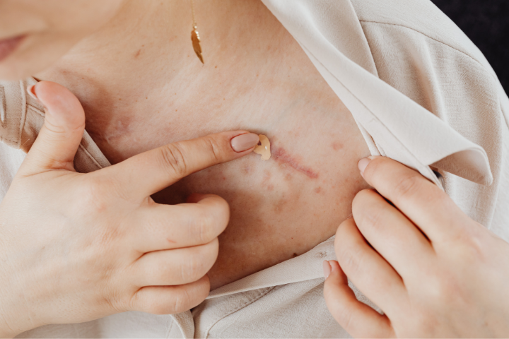
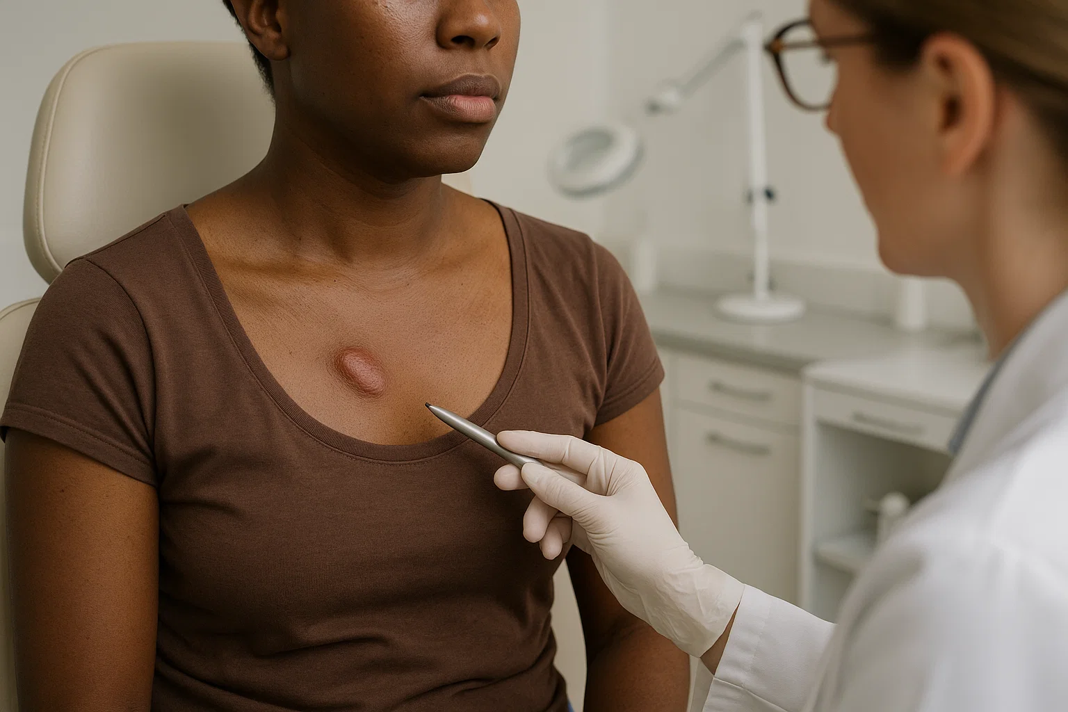
| Body Location | Skin Tension Level | Keloid Prevalence | Prevention Priority |
| Chest | Very High | 25-35% | Critical |
| Shoulders | High | 20-30% | High |
| Earlobes | Moderate | 15-25% | Moderate |
| Arms/Legs | Variable | 10-20% | Moderate |
| Face | Low | 5-10% | Standard |
Mechanosensitive Pathways in Keloids:
YAP/TAZ Signalling:
•🔧 Mechanosensitive transcription factors responding to tissue stiffness
•🔧 Overactivated in keloids due to increased tissue tension
•🔧 Drives proliferation and collagen production
•🔧 Creates feedback loop – stiffness promotes more stiffness
Ion Channel Dysfunction:
•🔧 Mechanosensitive ion channels detect physical forces
•🔧 Overactive in keloid fibroblasts causing excessive calcium influx
•🔧 Triggers proliferation signals in response to normal movement
•🔧 Explains keloid growth in high-movement areas
Genetic Factors in Keloid Susceptibility
Inheritance Patterns and Family Risk
Keloid Inheritance Characteristics:
Genetic Inheritance Pattern:
•👨👩👧👦 Autosomal dominant with incomplete penetrance
•👨👩👧👦 Variable expression even within families
•👨👩👧👦 Environmental triggers required for manifestation
•👨👩👧👦 Anticipation possible – earlier onset in subsequent generations
Family Risk Assessment:
| Family History | Keloid Risk | Genetic Testing Recommended |
| No family history | 1-3% | Not routinely needed |
| Distant relatives | 5-8% | Consider if other risk factors |
| Siblings affected | 15-25% | Recommended |
| Parents affected | 20-35% | Strongly recommended |
| Multiple relatives | 35-50% | Essential |
Specific Genetic Markers Identified in 2025
Major Keloid Susceptibility Genes:
NEDD4 Gene Variants:
•🧬 Function: Protein degradation and cell signalling
•🧬 Keloid association: 3.2x increased risk with specific variants
•🧬 Mechanism: Impaired protein turnover in fibroblasts
•🧬 Population frequency: 15% in African populations, 3% in European
FOXL2 Gene Polymorphisms:
•🧬 Function: Transcription factor regulating cell proliferation
•🧬 Keloid association: 2.8x higher susceptibility
•🧬 Mechanism: Dysregulated cell cycle control
•🧬 Clinical relevance: Predicts treatment response
PAI-1 Gene Mutations:
•🧬 Function: Controls extracellular matrix breakdown
•🧬 Keloid association: 4.1x elevated risk
•🧬 Mechanism: Impaired scar remodelling
•🧬 Treatment implications: Affects therapy selection
HLA Allele Associations:
| HLA Allele | Population Frequency | Keloid Risk Increase | Mechanism |
| HLA-DRB1*15 | 12% (European), 25% (African) | 3.5x | Immune dysregulation |
| HLA-DQB1*06 | 8% (European), 18% (African) | 2.8x | Inflammatory response |
| HLA-B*53 | 2% (European), 15% (African) | 4.2x | Antigen presentation |
Epigenetic Modifications in Keloids
DNA Methylation Patterns:
Hypermethylated Genes in Keloids:
•🔬 CDKN2A – Cell cycle control gene silenced
•🔬 TIMP3 – Matrix remodelling inhibitor reduced
•🔬 SFRP1 – Wnt signalling inhibitor suppressed
Hypomethylated Genes in Keloids:
•🔬 COL1A1 – Collagen production gene overexpressed
•🔬 ACTA2 – Smooth muscle actin gene activated
•🔬 TGFB1 – TGF-β gene overexpressed
MicroRNA Dysregulation:
| MicroRNA | Normal Function | Keloid Expression | Effect |
| miR-29 | Collagen regulation | 60% reduced | Excessive collagen |
| miR-200 | EMT prevention | 70% reduced | Fibroblast activation |
| miR-21 | Proliferation control | 5x increased | Hyperproliferation |
| miR-199 | Angiogenesis regulation | 3x increased | Excessive blood vessels |
Environmental Triggers and Risk Factors
Wound Characteristics That Promote Keloids
High-Risk Wound Features:
| Wound Characteristic | Low Risk | Moderate Risk | High Risk |
| Depth | Superficial (epidermis only) | Partial thickness | Full thickness |
| Size | <1cm | 1-3cm | >3cm |
| Location | Low-tension areas | Moderate tension | High-tension areas |
| Healing time | <2 weeks | 2-4 weeks | >4 weeks |
| Infection | None | Mild, brief | Severe, prolonged |
Procedure-Related Risk Factors:
Surgical Procedures by Risk Level:
High-Risk Procedures (>30% keloid rate in susceptible individuals):
•🔴 Cardiac surgery – Chest incisions in high-tension area
•🔴 Breast surgery – High-tension location, large incisions
•🔴 Ear surgery – Cartilage involvement, poor blood supply
•🔴 Burn reconstruction – Large areas, multiple procedures
Moderate-Risk Procedures (15-30% keloid rate):
•🟡 Abdominal surgery – Moderate tension, variable healing
•🟡 Orthopaedic surgery – Joint areas, movement stress
•🟡 Facial surgery – Variable location risk
•🟡 Cosmetic procedures – Elective, often multiple
Lower-Risk Procedures (<15% keloid rate):
•🟢 Minor skin procedures – Small, low-tension areas
•🟢 Endoscopic surgery – Minimal incisions
•🟢 Laser procedures – Controlled tissue damage
•🟢 Injection procedures – Minimal trauma
Hormonal Influences on Keloid Formation
Age and Hormonal Status:
Keloid Incidence by Age Group:
| Age Group | Keloid Incidence | Hormonal Factors | Clinical Implications |
| 0-10 years | 2-5% | Growth hormones | Rare, usually genetic |
| 11-20 years | 15-25% | Puberty, sex hormones | Peak incidence period |
| 21-30 years | 10-20% | Reproductive years | Pregnancy effects |
| 31-40 years | 8-15% | Hormonal stability | Moderate risk |
| 40+ years | 5-10% | Declining hormones | Lower incidence |
Pregnancy and Keloid Formation:
Pregnancy-Related Changes:
•🤰 Oestrogen elevation – Stimulates collagen production
•🤰 Progesterone effects – Modulates inflammatory response
•🤰 Growth hormone increases – Promotes tissue growth
•🤰 Cortisol changes – Affects healing patterns
Clinical Observations:
•📊 30% of women report keloid changes during pregnancy
•📊 Existing keloids may grow or become more symptomatic
•📊 New keloids more likely to form from pregnancy-related procedures
•📊 Postpartum period shows increased keloid activity
Thyroid Hormone Effects:
| Thyroid Status | Keloid Risk | Mechanism | Clinical Management |
| Hyperthyroid | Increased | Accelerated metabolism, healing | Monitor closely |
| Hypothyroid | Variable | Impaired healing, inflammation | Optimise thyroid function |
| Normal | Baseline | Standard healing patterns | Routine care |
Environmental and Lifestyle Factors
Climate and Environmental Influences:
London-Specific Environmental Factors:
•🌧️ High humidity – May affect wound healing and keloid symptoms
•🌫️ Air pollution – Inflammatory triggers in urban environment
•☁️ Limited sunlight – Vitamin D deficiency affecting healing
•🌡️ Temperature fluctuations – Seasonal effects on keloid symptoms
Occupational and Lifestyle Risk Factors:
| Risk Factor | Mechanism | Keloid Impact | Prevention Strategy |
| Smoking | Impaired circulation, delayed healing | 2.5x increased risk | Cessation essential |
| Alcohol excess | Immune suppression, poor nutrition | 1.8x increased risk | Moderation recommended |
| Chronic stress | Cortisol elevation, immune dysfunction | 2.1x increased risk | Stress management |
| Poor nutrition | Impaired healing capacity | 1.6x increased risk | Nutritional optimisation |
The Keloid Microenvironment: A Complex Ecosystem
Cellular Composition of Keloid Tissue
Cell Population Analysis:
| Cell Type | Normal Scar | Keloid Tissue | Functional Role |
| Fibroblasts | 60% | 45% | Collagen production |
| Myofibroblasts | 15% | 35% | Contraction, tension |
| Endothelial cells | 10% | 12% | Blood vessel formation |
| Immune cells | 10% | 15% | Inflammation |
| Other cells | 5% | 3% | Support functions |
Extracellular Matrix Abnormalities:
Collagen Composition Changes:
| Collagen Type | Normal Ratio | Keloid Ratio | Functional Impact |
| Type I | 80% | 65% | Reduced strength |
| Type III | 15% | 30% | Increased thickness |
| Type V | 3% | 4% | Altered structure |
| Type VI | 2% | 1% | Reduced flexibility |
Matrix Organisation:
•🔬 Normal scars: Parallel collagen fibres
•🔬 Keloids: Whorl-like, disorganised patterns
•🔬 Cross-linking: Excessive, abnormal patterns
•🔬 Proteoglycans: Altered composition and distribution
Vascular Abnormalities in Keloids
Blood Vessel Characteristics:
Keloid Vasculature Features:
•🩸 Increased vessel density – 2-3x more blood vessels
•🩸 Abnormal vessel structure – Dilated, tortuous patterns
•🩸 Endothelial dysfunction – Increased permeability
•🩸 Angiogenic factor elevation – VEGF, FGF overexpression
Hypoxia and Metabolic Changes:
| Parameter | Normal Tissue | Keloid Centre | Keloid Edge |
| Oxygen level | Normal | 40% reduced | 20% reduced |
| Glucose metabolism | Oxidative | Glycolytic | Mixed |
| Lactate production | Low | 5x elevated | 3x elevated |
| pH level | 7.4 | 7.1 | 7.2 |
Translating Science into Treatment: 2025 Advances
Targeted Molecular Therapies Based on Pathogenesis
Pathway-Specific Treatments:
TGF-β Pathway Modulators:
•💊 Pirfenidone – Reduces TGF-β signalling, 45% improvement
•💊 Nintedanib – Multi-kinase inhibitor, 38% success rate
•💊 Anti-TGF-β antibodies – Direct pathway blocking, clinical trials
JAK/STAT Inhibitors:
•💊 Topical JAK inhibitors – Reduce inflammation, 52% improvement
•💊 Oral JAK inhibitors – Systemic effects, severe cases only
•💊 Combination protocols – Enhanced effectiveness
Hedgehog Pathway Targeting:
•💊 HHIP replacement therapy – Restore normal signalling
•💊 GLI inhibitors – Block downstream effects
•💊 SMO antagonists – Prevent pathway activation
Personalised Treatment Based on Genetic Profiling
Genetic Testing for Treatment Selection:
| Genetic Profile | Recommended Treatment | Expected Response | Alternative Options |
| NEDD4 variants | Proteasome modulators | 70% improvement | JAK inhibitors |
| FOXL2 mutations | Cell cycle inhibitors | 65% improvement | TGF-β blockers |
| PAI-1 deficiency | Matrix modulators | 75% improvement | Combination therapy |
| HLA high-risk | Immunomodulators | 60% improvement | Anti-inflammatory |
Precision Medicine Approach:
Treatment Algorithm Based on Keloid Characteristics:
1.Genetic testing – Identify susceptibility markers
2.Molecular profiling – Analyse keloid tissue characteristics
3.Pathway assessment – Determine dominant mechanisms
4.Treatment selection – Choose targeted therapies
5.Response monitoring – Adjust protocols based on outcomes
Prevention Strategies Informed by Science
Mechanism-Based Prevention:
Targeting Inflammation:
•🛡️ Early anti-inflammatory protocols – Prevent chronic inflammation
•🛡️ Mast cell stabilisers – Reduce inflammatory mediator release
•🛡️ Cytokine modulators – Balance inflammatory response
Addressing Mechanical Factors:
•🛡️ Tension-reduction techniques – Minimise mechanotransduction
•🛡️ Pressure therapy – Counteract tissue tension
•🛡️ Movement protocols – Optimise healing mechanics
Genetic Risk Management:
•🛡️ High-risk identification – Genetic screening programs
•🛡️ Prophylactic treatments – Prevent keloid formation
•🛡️ Family counselling – Genetic risk education
Future Directions: The Next Frontier in Keloid Science
Emerging Research Areas
Single-Cell Technologies:
•🔬 Spatial transcriptomics – Map gene expression in tissue context
•🔬 Single-cell proteomics – Understand protein function
•🔬 Epigenetic mapping – Identify regulatory mechanisms
•🔬 Metabolomics – Analyse cellular metabolism
Artificial Intelligence Applications:
•🤖 Predictive modelling – Forecast keloid development
•🤖 Treatment optimisation – Personalise therapy selection
•🤖 Drug discovery – Identify new therapeutic targets
•🤖 Outcome prediction – Estimate treatment success
Regenerative Medicine:
•🧪 Stem cell therapies – Restore normal healing
•🧪 Tissue engineering – Create normal scar tissue
•🧪 Gene editing – Correct genetic defects
•🧪 Exosome therapy – Deliver healing signals
Frequently Asked Questions (FAQs)
Basic Science Questions
Why do some people get keloids while others don’t?
Keloid formation results from a combination of genetic susceptibility (inherited risk factors), environmental triggers (injuries, procedures), and individual healing responses. About 15-20% of people of African descent and 5-8% of Asian populations have genetic predisposition, but environmental factors determine whether keloids actually develop.
Are keloids a type of cancer?
No, keloids are benign (non-cancerous) growths. While they share some characteristics with tumours (excessive cell growth, resistance to cell death), they don’t spread to other parts of the body and aren’t life-threatening. However, they can be persistent and challenging to treat.
Can keloids turn into cancer?
Keloids themselves don’t become cancerous. However, very rarely, aggressive treatments or chronic irritation might contribute to skin cancer development. This is why proper medical management is important rather than attempting self-treatment.
Why do keloids itch and hurt?
Keloids contain 10-15 times more mast cells than normal tissue. These cells release histamine and other inflammatory mediators that cause itching. Pain results from nerve compression by the thick scar tissue and ongoing inflammation.
Genetic and Risk Factor Questions
If my parent has keloids, will I definitely get them?
Not necessarily. Keloid susceptibility follows an autosomal dominant pattern with incomplete penetrance, meaning you have a 20-35% chance of developing keloids if a parent is affected. Environmental triggers (injuries, procedures) are still required for keloids to actually form.
Can genetic testing predict if I’ll get keloids?
Genetic testing can identify increased susceptibility but can’t predict with certainty whether you’ll develop keloids. Testing is most useful for high-risk individuals planning procedures, allowing for enhanced prevention protocols.
Do keloids affect all ethnicities equally?
No, there are significant ethnic differences. African and Afro-Caribbean populations have 15-20% prevalence, Asian populations 8-12%, Hispanic/Latino 6-10%, and Caucasian populations 1-3%. These differences are primarily genetic.
Why are keloids more common in certain body areas?
Keloids preferentially form in high-tension areas where mechanical forces are greatest. The chest, shoulders, and earlobes experience more skin tension, which activates mechanotransduction pathways that promote keloid formation.
Treatment and Prevention Questions
How does understanding keloid science help with treatment?
Scientific understanding has led to targeted therapies that address specific molecular mechanisms. For example, knowing that TGF-β signalling is overactive has led to TGF-β inhibitors, while understanding genetic factors allows for personalised treatment selection.
Can keloids be prevented if I know I’m at risk?
Yes, understanding your risk allows for implementation of prevention protocols that can reduce keloid formation by up to 78%. These include genetic testing, enhanced wound care, early intervention, and avoiding high-risk procedures when possible.
Why do some treatments work for some people but not others?
Keloids result from different molecular mechanisms in different individuals. Genetic variations, specific pathway abnormalities, and environmental factors all influence treatment response. This is why personalised medicine approaches are becoming more important.
Will understanding keloid science lead to better treatments?
Absolutely. The molecular insights gained in 2025 have already led to breakthrough treatments like the ASAP Protocol and targeted molecular therapies. Continued research into keloid mechanisms will likely yield even more effective treatments.
London-Specific Questions
Are there environmental factors in London that affect keloid formation?
London’s urban environment includes factors that may influence keloid development, including air pollution (inflammatory triggers), high humidity (affecting wound healing), and limited sunlight (vitamin D deficiency). However, these are manageable with proper care.
Where can I access genetic testing for keloid risk in London?
Genetic testing for keloid susceptibility is available at London Keloid Scar Clinic and select other centres. The test involves a simple saliva sample and provides comprehensive risk assessment within 7-10 days.
How does the latest keloid science research apply to treatments available in London?
London Keloid Scar Clinic incorporates the latest scientific findings into treatment protocols, including molecular targeted therapies, genetic testing for personalised treatment, and AI-assisted treatment planning based on current research.
Can I participate in keloid research studies in London?
Yes, several London centres conduct keloid research studies. Participation can provide access to cutting-edge treatments while contributing to scientific advancement. Contact London Keloid Scar Clinic for information about current studies.
Conclusion: Knowledge Empowers Better Outcomes
Understanding the complex science behind keloid formation provides the foundation for effective prevention, early intervention, and treatment. The remarkable advances in keloid research during 2025 have transformed our approach to these challenging scars, offering new hope through targeted, scientifically-informed therapies.
Key Scientific Insights:
•🧬 Genetic factors create susceptibility but don’t guarantee keloid formation
•🔬 Molecular mechanisms can be targeted with specific therapies
•📊 Environmental triggers can be identified and managed
•🎯 Personalised approaches based on individual characteristics improve outcomes
•💡 Prevention strategies informed by science are highly effective
How This Knowledge Helps You:
For Prevention:
•Risk assessment using genetic and clinical factors
•Targeted prevention protocols based on your specific risk profile
•Early intervention when abnormal healing is detected
•Lifestyle modifications to optimise healing capacity
For Treatment:
•Personalised therapy selection based on keloid characteristics
•Combination approaches targeting multiple pathways
•Monitoring protocols to optimise treatment response
•Long-term management strategies for sustained success
Ready to Apply This Knowledge?
Book your consultation at London Keloid Scar Clinic to:
•Receive genetic risk assessment with latest testing technology
•Understand your specific keloid mechanisms through advanced analysis
•Access science-based treatments tailored to your needs
•Develop personalised prevention or treatment protocols
Contact Information:
•Website: londonkeloidscarclinic.co.uk
•Scientific Consultation: Available for complex cases
•Location: Central London with excellent transport links
2nd floor, 4 Harley St, London W1G 9QY, United Kingdom
Understanding your keloids at the molecular level empowers you to make informed decisions about prevention and treatment. The scientific advances of 2025 offer unprecedented opportunities for successful keloid management—take advantage of this knowledge to achieve the best possible outcomes.
This article was written by the clinical team at London Keloid Scar Clinic. Information is based on the latest scientific research and clinical understanding. Individual results may vary. Consult with qualified medical professionals for personalised advice.